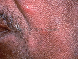
Kimura disease, also known as eosinophilic hyperplastic lymphogranuloma or atypical granulation associated with hyperplastic abnormalities in the lymphoid tissue, is a rare, chronic inflammatory disorder of unknown etiology. It is characterized by the development of painless cervical lymphadenopathy, peripheral eosinophilia, elevated serum immunoglobulin E (IgE) levels, and/or subcutaneous lymphoid masses, usually on the head and neck.
Kimura disease is most common in Asia. Patients with Kimura disease are usually young adults, although the disease may present during adolescence, particularly in East and Southeast Asian males. Men are affected more often than women. About 15%-20% of patients with Kimura disease also have nephrotic syndrome, although the pathophysiologic basis for this association is unclear, and acute glomerulonephritis may also occur. Kimura disease may also result in a hypercoagulable sequela, thought to be related to hypereosinophilia and inflammation, that can cause visible ischemia of the extremities or visceral venous thromboembolism.
It is thought that the disease may represent a hypersensitivity or autoimmune process, but regardless, the end result is clearly an abnormal proliferation of lymphoid follicles and vascular endothelium. Of note, angiolymphoid hyperplasia with eosinophilia (ALHE) and Kimura disease, once thought to be disease variants, are now recognized to be separate, distinct diseases with specific clinical and histologic features.
Kimura disease
Alerts and Notices
Important News & Links
Synopsis

Codes
ICD10CM:
D21.9 – Benign neoplasm of connective and other soft tissue, unspecified
SNOMEDCT:
399894006 – Kimura disease
D21.9 – Benign neoplasm of connective and other soft tissue, unspecified
SNOMEDCT:
399894006 – Kimura disease
Look For
Subscription Required
Diagnostic Pearls
Subscription Required
Differential Diagnosis & Pitfalls

To perform a comparison, select diagnoses from the classic differential
Subscription Required
Best Tests
Subscription Required
Management Pearls
Subscription Required
Therapy
Subscription Required
References
Subscription Required
Last Reviewed:03/22/2026
Last Updated:03/23/2026
Last Updated:03/23/2026
Kimura disease