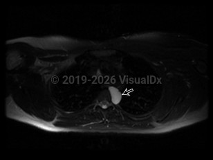
Bronchogenic cyst in Adult
Alerts and Notices
Important News & Links
Synopsis

A bronchogenic cyst is a congenital respiratory tract malformation arising as an embryonic out-pouching of the foregut or trachea. Bronchogenic cysts can be found anywhere along the tracheobronchial tree and are often asymptomatic unless they become infected. Cysts typically appear as sharply marginated, water-density lesions on chest x-ray and may contain air-fluid levels when infected.
Symptoms can vary, but patients will present with failure to thrive or with airway obstruction. Failure to thrive is likely due to dysphagia and subsequent poor oral nutrition intake. Hemoptysis can occur when the bronchogenic cyst erodes into surrounding vasculature.
Symptoms can vary, but patients will present with failure to thrive or with airway obstruction. Failure to thrive is likely due to dysphagia and subsequent poor oral nutrition intake. Hemoptysis can occur when the bronchogenic cyst erodes into surrounding vasculature.
Codes
ICD10CM:
J98.4 – Other disorders of lung
SNOMEDCT:
762195006 – Congenital bronchogenic cyst
J98.4 – Other disorders of lung
SNOMEDCT:
762195006 – Congenital bronchogenic cyst
Look For
Subscription Required
Diagnostic Pearls
Subscription Required
Differential Diagnosis & Pitfalls

To perform a comparison, select diagnoses from the classic differential
Subscription Required
Best Tests
Subscription Required
Management Pearls
Subscription Required
Therapy
Subscription Required
References
Subscription Required
Last Reviewed:03/21/2019
Last Updated:05/05/2019
Last Updated:05/05/2019
Bronchogenic cyst in Adult

