Oral leukemic infiltration (OLI), also known as oral malignant infiltration (OMI), is the presence of localized or generalized tissue hyperplasia of the mouth caused by collections of malignant white blood cells (WBCs) or their precursors. OLI may occur with virtually any type of leukemia; however, it is most commonly seen with acute leukemias, such as acute myeloid leukemia and acute lymphoblastic leukemia.
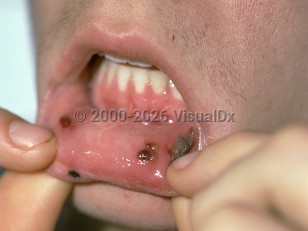
Lesions may vary in distribution and morphology, and may include gingival hyperplasia, petechiae, oral ulcerations, and mucosal pallor. Patients may endorse gingival bleeding and oral or laryngeal pain. Due to leukemia-related and treatment-related immunosuppression, lesions may become secondarily infected by bacterial, viral, yeast, or fungal pathogens.
Oral lesions of leukemia may develop as the initial presenting sign of leukemia or may occur with disease relapse. Concomitant lymphadenopathy may occur, and patients may endorse B symptoms (symptoms associated with lymphoma) such as fever, night sweats, fatigue, unintentional weight loss, and loss of appetite.
All patient populations with leukemia may be at risk for OLI; however, the highest incidence tends to be in adults aged 40-49 years with acute myeloid leukemia. OLI has a slight male predominance.
Importantly, OLI often portends a poor prognosis and has been associated with worse outcomes. Survival rates are typically low for those affected by OLI, with one study noting only 14.3% survival at 21 months. Myeloid sarcoma (chloroma), an extramedullary tumor of acute myeloid leukemia, may present with oral involvement and is associated with a more aggressive leukemia and high risk of mortality.
Oral leukemic infiltration - Oral Mucosal Lesion
Alerts and Notices
Important News & Links
Synopsis

Codes
ICD10CM:
C95.90 – Leukemia, unspecified not having achieved remission
SNOMEDCT:
404090003 – Malignant infiltration of oral cavity by underlying tumor
446688004 – Leukemic infiltration
C95.90 – Leukemia, unspecified not having achieved remission
SNOMEDCT:
404090003 – Malignant infiltration of oral cavity by underlying tumor
446688004 – Leukemic infiltration
Look For
Subscription Required
Diagnostic Pearls
Subscription Required
Differential Diagnosis & Pitfalls

To perform a comparison, select diagnoses from the classic differential
Subscription Required
Best Tests
Subscription Required
Management Pearls
Subscription Required
Therapy
Subscription Required
References
Subscription Required
Last Reviewed:12/14/2025
Last Updated:12/14/2025
Last Updated:12/14/2025
Oral leukemic infiltration - Oral Mucosal Lesion