Contents

SynopsisCodesLook ForDiagnostic PearlsDifferential Diagnosis & PitfallsBest TestsManagement PearlsTherapyDrug Reaction DataReferences

View all Images (23)

Photoonycholysis - Nail and Distal Digit
Other Resources UpToDate PubMed

Photoonycholysis - Nail and Distal Digit

Contributors: Shari Lipner MD, PhD, Susan Burgin MD, Bertrand Richert MD, Robert Baran MD
Other Resources UpToDate PubMed

Synopsis

Photoonycholysis refers to separation of the nail plate from the nail bed induced by ultraviolet light following recent therapy with medications or in the setting of a photosensitive blistering disorder, such as the porphyrias or pseudoporphyria.

Common causative medications include tetracycline derivatives, psoralens, aminolevulinic acid, griseofulvin, oral contraceptives, fluoroquinolones, voriconazole, or chloramphenicol. Photoonycholysis usually appears more than 2 weeks after exposure to the drug. It may occur as part of Segal's triad of photosensitivity of the skin, discoloration of the nails, and onycholysis. The latter, however, may appear independently in the absence of photosensitive reaction elsewhere. Pseudoporphyria is also usually drug-induced. While commonly associated with NSAIDs, antibiotics, and diuretics with sulfa moieties, it can also be seen in chronic renal failure (with or without hemodialysis) and with UVA exposure such as tanning beds, psoralen plus UVA (PUVA) therapy, and excessive natural sun.

There are 4 types of photoonycholysis:
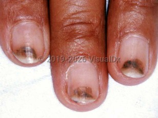
  • Several fingers are involved. The separating portion of the nail plate is half-moon shaped and concave distally with pigmentation of variable intensity and a well-demarcated proximal border.
  • Only one finger is affected. There is a well-defined circular notch, which opens distally and has a brownish hue proximally.
  • It occurs in the central part of the pink nail bed on several fingers. It is initially a round, yellow staining that turns red after 5-10 days.
  • It is characterized by bullae under the nails with associated photoonycholysis due to tetracycline as well as in porphyria cutanea tarda, erythropoietic porphyria, erythropoietic protoporphyria, variegata porphyria, and in pseudoporphyria.
There is no relationship between etiology and the different clinical varieties of photoonycholysis.

Photoonycholysis may also occur in children, Common culprits include tetracycline derivatives, griseofulvin, and voriconazole. In absence of an obvious medication, a workup for systemic causes including porphyrias should be performed.

Related topics: Onycholysis, Pseudoporphyria, Porphyria cutanea tarda

Codes

ICD10CM:
L56.8 – Other specified acute skin changes due to ultraviolet radiation

SNOMEDCT:
95342006 – Photoonycholysis

Look For

Subscription Required

Diagnostic Pearls

Subscription Required

Differential Diagnosis & Pitfalls

To perform a comparison, select diagnoses from the classic differential

Subscription Required

Best Tests

Subscription Required

Management Pearls

Subscription Required

Therapy

Subscription Required

Drug Reaction Data

Subscription Required

References

Subscription Required

Last Reviewed:06/05/2018
Last Updated:06/05/2018
Copyright © 2025 VisualDx®. All rights reserved.
Photoonycholysis - Nail and Distal Digit
A medical illustration showing key findings of Photoonycholysis : Nail color change, Nail pain, Sun exposure - recent intense, Onycholysis
Clinical image of Photoonycholysis - imageId=1659036. Click to open in gallery.  caption: 'Half-moon-shaped, red, purple-black, and brown subungual discoloration of the distal fingernails.'
Half-moon-shaped, red, purple-black, and brown subungual discoloration of the distal fingernails.
Copyright © 2025 VisualDx®. All rights reserved.