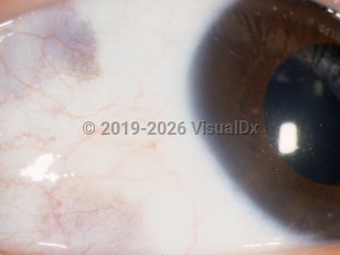
Conjunctival melanosis - External and Internal Eye
Alerts and Notices
Important News & Links
Synopsis

Primary acquired melanosis (PAM) of the conjunctiva presents as a unilateral, flat, patchy, non-cystic, and brown-pigmented lesion of the conjunctival epithelium. It is seen mostly in middle-aged, fair-skinned, and older, white patients. It is caused by the proliferation of atypical melanocytes in the epithelium that do not invade the subepithelial tissue unless there has been malignant transformation. Since it can occur in any part of the conjunctiva, it is important to examine the underside of the eyelid. This lesion can remain dormant for years or show slow progression. It is the most important precursor of conjunctival malignant melanoma with some 1–30% of cases following that course. There are no associated symptoms.
Codes
ICD10CM:
H11.139 – Conjunctival pigmentations, unspecified eye
SNOMEDCT:
91268007 – Conjunctival melanosis
H11.139 – Conjunctival pigmentations, unspecified eye
SNOMEDCT:
91268007 – Conjunctival melanosis
Look For
Subscription Required
Diagnostic Pearls
Subscription Required
Differential Diagnosis & Pitfalls

To perform a comparison, select diagnoses from the classic differential
Subscription Required
Best Tests
Subscription Required
Management Pearls
Subscription Required
Therapy
Subscription Required
References
Subscription Required
Last Updated:07/02/2007

