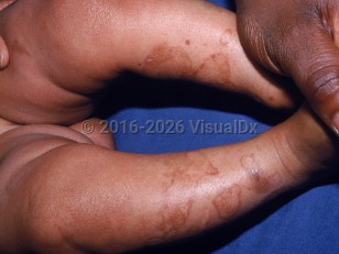
Kindler syndrome in Infant/Neonate
Alerts and Notices
Important News & Links
Synopsis

Kindler syndrome (KS) is a rare type of inherited epidermolysis bullosa (EB), which is a group of mechanobullous disorders characterized by defects in the basement membrane zone (BMZ). Instability of the BMZ manifests clinically as skin fragility and bulla formation at sites of trauma.
KS is inherited in an autosomal recessive pattern that results from a loss-of-function in the FERMT1 gene (formerly KIND1), which encodes a protein known as fermitin family homolog 1 (FFH1), a focal adhesion protein that links the actin cytoskeleton with the underlying extra cellular matrix. Weakness at this point causes splits at various levels, most often above the basal keratinocytes.
Like other forms of EB, acral blisters in KS are typically noted soon after birth or in early infancy. Disease severity may range from mild, with minimal skin disease, to severe, with involvement of mucosal sites (such as conjunctiva, vagina, urethra, anus, and mouth) and gastrointestinal tract (causing esophageal strictures and colitis). Patients with mild disease may not be diagnosed until late adulthood.
Features that distinguish KS from other types of EB are photosensitivity (worst in childhood), progressive poikiloderma, and marked skin atrophy, which begin early in the disease course. While blistering and photosensitivity tend to diminish in severity with age, poikiloderma is permanent. Scarring is usually minimal, but repeated trauma can result in development of scar tissue.
Other clinical findings include mucosal involvement, including gingivitis, mucositis (hemorrhagic), labial leukokeratosis, periodontal disease / periodontitis, tooth enamel defects, and loss of teeth. Palmoplantar hyperkeratosis, nail dystrophy, and pseudosyndactyly can also be seen, as well as loss of fingerprints. Ocular involvement can cause ectropion. Genital involvement can cause urethral stenosis and phimosis. Joint laxity and esophageal stenosis may also be seen.
Long-term complications include the development of aggressive squamous cell carcinoma (SCC). SCC is also reported on acral skin and the mouth.
Related topics: dystrophic EB, EB acquisita, EB simplex, generalized severe EB simplex, junctional EB
Codes
ICD10CM:
Q81.8 – Other epidermolysis bullosa
Q82.8 – Other specified congenital malformations of skin
SNOMEDCT:
238836000 – Kindler's syndrome
Q81.8 – Other epidermolysis bullosa
Q82.8 – Other specified congenital malformations of skin
SNOMEDCT:
238836000 – Kindler's syndrome
Look For
Subscription Required
Diagnostic Pearls
Subscription Required
Differential Diagnosis & Pitfalls

To perform a comparison, select diagnoses from the classic differential
Subscription Required
Best Tests
Subscription Required
Management Pearls
Subscription Required
Therapy
Subscription Required
References
Subscription Required
Last Reviewed:05/07/2024
Last Updated:05/21/2024
Last Updated:05/21/2024
Kindler syndrome in Infant/Neonate

