Contents

SynopsisCodesLook ForDiagnostic PearlsDifferential Diagnosis & PitfallsBest TestsManagement PearlsTherapyDrug Reaction DataReferences

View all Images (116)

Other Resources UpToDate PubMed

Vasculitis

See also in: Cellulitis DDx,External and Internal Eye
Contributors: Noah Craft MD, PhD, Lindy P. Fox MD, Jeffrey D. Bernhard MD, Michael D. Tharp MD, Art Papier MD, Neil Shear MD, Lowell A. Goldsmith MD, MPH
Other Resources UpToDate PubMed

Synopsis

Vasculitis is a nonspecific term that encompasses a large and heterogeneous group of disorders that are characterized by inflammation of blood vessels. No uniform classification system for vasculitis exists, although subtypes are often categorized by the size of the vessels involved, the type of circulating immune complexes, and other histopathologic and clinical features. In many cases, damage to the blood vessels results in leakage of red blood cells and the development of palpable purpura on the skin. Other common cutaneous signs of vasculitis include skin ulcerations, Raynaud phenomenon, and livedo reticularis.

It is important to distinguish a primary systemic vasculitis from one associated with medications, infection, malignancy, or a connective tissue disorder, as the best course of treatment may differ. Drug-induced vasculitis should be quickly recognized and managed by removing the causative medication. Ongoing drug-induced vasculitis can lead to glomerulonephritis, pulmonary hemorrhage, mononeuritis multiplex, and other complications of end-organ involvement.

Several types of vasculitis are more likely than others to be mistaken for cellulitis. Among these are polyarteritis nodosa and variants of leukocytoclastic (hypersensitivity) vasculitis such as erythema elevatum diutinum and urticarial vasculitis. Kawasaki disease and immunoglobulin A vasculitis (formerly Henoch-Schönlein purpura) may present with some lesions that mimic cellulitis, but these diseases occur predominantly in children. On rarer occasions, other vasculitides such as granulomatosis with polyangiitis, Behçet syndrome, cryoglobulinemia, and livedoid vasculopathy may present similarly to cellulitis. Other primary vasculitic syndromes include eosinophilic granulomatosis with polyangiitis, microscopic polyangiitis, Takayasu arteritis, and giant cell (temporal) arteritis. Vasculitis may occur in association with such diseases as rheumatoid arthritis, systemic lupus erythematosus, and dermatomyositis.

Polyarteritis nodosa (PAN) refers to a necrotizing vasculitis of small- and medium-sized arterioles. It is the least distinctive of the vasculitides. The exact etiology is unknown, but it likely involves immune complex deposition, autoantibodies, inflammatory mediators, and adhesion molecules. Any organ may be affected, but PAN most commonly involves the skin, peripheral nerves, kidneys, joints, and gastrointestinal (GI) tract. Symptoms may include malaise, fever (in 50% of cases), weakness, myalgias, arthralgias, abdominal pain, cutaneous ulcers, livedo reticularis, testicular pain, and weight loss. Polyarteritis nodosa has been associated with infection with hepatitis B, hepatitis C, human immunodeficiency virus (HIV), cytomegalovirus (CMV), parvovirus B19, human T-cell lymphotropic virus (HTLV), and streptococci. There appears to be an association with inflammatory bowel disease. Polyarteritis nodosa usually affects individuals in mid- to late adulthood. It is slightly more common in men, and there is no apparent racial predilection.

"Leukocytoclastic vasculitis" (LCV) is a pathologic term that refers to inflammation of small blood vessels, usually with an inflammatory infiltrate and necrosis of the vessel wall. This form of vasculitis may be localized to the skin where it is most commonly seen as palpable purpura, or it may also involve the other organs such as the kidneys, the joints, or the GI tract. The exact pathogenesis remains elusive, but LCV appears to involve circulating immune complexes, autoantibodies, and inflammatory mediators.

Erythema elevatum diutinum refers to a rare subtype of LCV. It is associated with upper respiratory infections (especially streptococcal), HIV, hematologic disease, and rheumatologic disorders. It presents with arthralgias and erythematous to violaceous papules, plaques, and nodules with a predilection for extensor surfaces.

Urticarial vasculitis is also considered to be a variant of LCV. This presentation may be idiopathic, or it can occur in association with serum sickness, connective tissue disorders, infections, and after the administration of potassium iodide or nonsteroidal anti-inflammatory agents (NSAIDs). Urticarial vasculitis affects mainly women. The skin lesions last 3-5 days. Episodic arthralgias are a major clinical manifestation and affect the wrists, fingers, knees, ankles, and toes. General features include fever, malaise, myalgias, lymphadenopathy, and hepatosplenomegaly.

Patients with antineutrophil cytoplasmic antibody (ANCA)-positive drug-induced vasculitis commonly have antibodies to multiple granule proteins, such as myeloperoxidase (MPO), cathepsin G, lactoferrin, and human leukocyte elastase (HLE).

Drug-induced vasculitis frequently presents with skin involvement and, if progressive, can include any of the following symptoms of systemic involvement: myalgias, arthralgias, dyspnea, hoarseness, vision impairment from retinal vessel involvement, and abdominal pain. Onset of the vasculitis is variable, ranging from weeks to years after starting therapy. The following medications are among the most frequent causes of drug-induced vasculitis: anti-thyroid agents such as propylthiouracil and methimazole, minocycline, hydralazine, phenytoin, granulocyte colony-stimulating factor (G-CSF), allopurinol, D-penicillamine, cefaclor, and methotrexate.

Methyldopa can cause life-threatening cardiac complications including cardiac myocyte necrosis and cardiomyopathy. Isotretinoin has been reported to induce vasculitis, sometimes with renal involvement. Levamisole, a veterinary medication banned for human use, has been shown to cause vasculitis. Cocaine contaminated with levamisole is a near-epidemic occurrence in cocaine users, leading to cocaine levamisole toxicity.

Unlike cellulitis, the lesions of vasculitis are often multifocal and/or bilateral. Ulceration, palpable purpura, and livedo reticularis may be present. Be sure to ask the patient about the presence of specific symptoms such as myalgias, arthralgias, hematuria, or neuropathy.

Codes

ICD10CM:
L95.9 – Vasculitis limited to the skin, unspecified

SNOMEDCT:
31996006 – Vasculitis

Look For

Subscription Required

Diagnostic Pearls

Subscription Required

Differential Diagnosis & Pitfalls

To perform a comparison, select diagnoses from the classic differential

Subscription Required

Best Tests

Subscription Required

Management Pearls

Subscription Required

Therapy

Subscription Required

Drug Reaction Data

Subscription Required

References

Subscription Required

Last Updated:03/12/2024
Copyright © 2025 VisualDx®. All rights reserved.
Vasculitis
See also in: Cellulitis DDx,External and Internal Eye
A medical illustration showing key findings of Vasculitis : Malaise, p-ANCA positive, Palpable purpura, Symmetric extremities distribution, Arthralgia, Myalgia, CRP elevated, ESR elevated
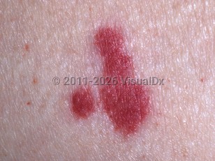
Clinical image of Vasculitis - imageId=323385. Click to open in gallery.  caption: 'A close-up of palpable purpura.'
A close-up of palpable purpura.
Copyright © 2025 VisualDx®. All rights reserved.