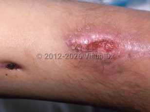
Cutaneous nocardiosis
Alerts and Notices
Important News & Links
Synopsis

Nocardia spp. are soil-dwelling, ubiquitous, gram-positive, filamentous, branching bacteria. The most common manifestation of nocardiosis is the pulmonary form, which typically affects patients with structural lung disease who have a malignancy or who are immunosuppressed.
Primary cutaneous nocardiosis (PCN) occurs after trauma (often with soil contact) to the skin and/or skin appendages and most often affects healthy patients. Lesions may occur suddenly and rapidly progress, reactivate after several years of dormancy, or slowly expand over 10 years. Lymphocutaneous spread may also be seen. A third manifestation is mycetoma. Here, a localized infection, typically of the foot, causes sinus tract formation and draining granules. Underlying tissues may become secondarily involved. Rarely, PCN can disseminate to other organs, but this is usually in the setting of significant immunocompromise.
PCN affects males more frequently than females. Risk factors include soil or sand exposure, gardening, farming, superficial injury from domestic shrubbery, intra-articular steroid injections, insect bites, cat scratches, or trauma from motor vehicle accidents leading to abrasions (ie, "road rash").
PCN may comprise up to 5% of all cases of nocardiosis. Nocardia infections can be found worldwide, but mycetomas are found more commonly in subtropical and tropical climates. PCN is usually caused by Nocardia brasiliensis but may be caused by nearly any species of Nocardia.
Secondary cutaneous nocardiosis occurs in association with underlying systemic nocardiosis. Fistulae form from underlying infected tissues. Subcutaneous abscesses are another manifestation. In underlying pulmonary disease, lesions occur over the chest. They are more widespread in disseminated disease.
Codes
ICD10CM:
A43.1 – Cutaneous nocardiosis
SNOMEDCT:
64650008 – Cutaneous nocardiosis
A43.1 – Cutaneous nocardiosis
SNOMEDCT:
64650008 – Cutaneous nocardiosis
Look For
Subscription Required
Diagnostic Pearls
Subscription Required
Differential Diagnosis & Pitfalls

To perform a comparison, select diagnoses from the classic differential
Subscription Required
Best Tests
Subscription Required
Management Pearls
Subscription Required
Therapy
Subscription Required
References
Subscription Required
Last Reviewed:03/20/2017
Last Updated:03/20/2017
Last Updated:03/20/2017
Cutaneous nocardiosis

