Blue rubber bleb nevus syndrome (BRBNS) is characterized by venous malformations of the skin and gastrointestinal (GI) tract. The syndrome can also rarely involve other internal organs. These include the adrenal glands, kidney, heart, lungs, bladder, penis, thyroid, spleen, central nervous system, ocular orbits, muscle, and bone.
BRBNS is rare; only about 200 cases have been described in the literature to date. The cause is unknown. Most cases of BRBNS occur spontaneously, although there have been reports of autosomal dominantly inherited cases. Some evidence suggests that the mutation for some cases may occur on chromosome 9p. Most patients with BRBNS have double (cis) somatic mutations in the TEK gene, which encodes TIE2, an endothelial cell tyrosine kinase receptor.
BRBNS has been reported to occur more frequently in White individuals. It affects males and females equally. Blue rubber bleb nevi are often present at birth or in early childhood, but onset has been reported in adults. According to the largest case series to date of 44 patients, 25% of patients develop their first lesion between the ages of 0 and 6 months, 25% develop lesions between the ages of 7 months and 11 years, 34% develop lesions between the ages of 12 and 18 years, and 30% develop lesions older than age 18 years.
Cutaneous and mucosal lesions typically appear as soft blue or purple nodules, but they can vary in appearance and can range from small blue-black punctate papules to large, disfiguring tumors. Subcutaneous hyperkeratotic lesions can also be observed in the mucosal or palmoplantar surfaces. Cutaneous lesions can be painful in up to 40% of patients. Over time, they increase in size and number.
Half (50%) of patients have skin lesions preceding GI lesions. Usually, GI involvement becomes evident during young adulthood. GI venous malformations predominantly occur in the small intestines, but they can occur anywhere from the mouth to the anus. These lesions bleed easily and can result in occult blood loss and iron deficiency anemia. They can also lead to abdominal pain, intussusception, volvulus, infarction, and internal hemorrhage. Consumptive coagulopathy may also develop.
Involvement of other organs may result in menorrhagia, intermittent proptosis, hematuria, epistaxis, hemoptysis, hemothorax, hemopericardium, thrombotic complications, focal seizures, spasticity, ataxia, weakness, arthralgias, decreased range of motion, and deformity.
Morbidity and mortality depend on the extent of visceral involvement. Most patients live a normal life span. However, hemorrhage from the GI tract can be fatal. Lesions involving the bones and joints can be uncomfortable and disfiguring and, rarely, require amputation. Infrequently, lesions of the central nervous system can be fatal.
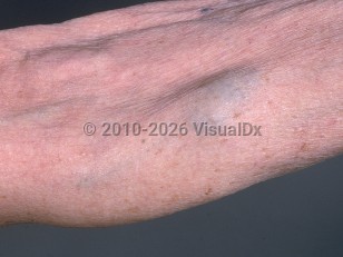
Blue rubber bleb nevus syndrome in Child
Alerts and Notices
Important News & Links
Synopsis

Codes
ICD10CM:
D18.00 – Hemangioma unspecified site
Q27.9 – Congenital malformation of peripheral vascular system, unspecified
SNOMEDCT:
254784002 – Blue rubber bleb nevus
D18.00 – Hemangioma unspecified site
Q27.9 – Congenital malformation of peripheral vascular system, unspecified
SNOMEDCT:
254784002 – Blue rubber bleb nevus
Look For
Subscription Required
Diagnostic Pearls
Subscription Required
Differential Diagnosis & Pitfalls

To perform a comparison, select diagnoses from the classic differential
Subscription Required
Best Tests
Subscription Required
Management Pearls
Subscription Required
Therapy
Subscription Required
References
Subscription Required
Last Reviewed:01/19/2026
Last Updated:02/02/2026
Last Updated:02/02/2026
Blue rubber bleb nevus syndrome in Child