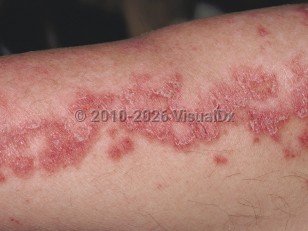
Subacute cutaneous lupus erythematosus (SCLE) is a subtype of cutaneous lupus that is characterized by smooth or scaly annular plaques with a predilection for photoexposed areas. The sides of the face, the lower neck, the upper trunk, and the extensor surfaces of the arms are the most commonly affected sites. Although scarring is not a characteristic finding, dyspigmentation is a common sequela.
SCLE prevalence ranges from 17-48 cases per 100 000 people. While the etiology remains poorly understood, there is a strong association with anti-Ro/SSA antibodies and SCLE. It is hypothesized that a complex interplay between genetic proclivity and environmental influences leads to a perpetuated autoimmune response. In addition, specific HLA types (1, B8, DR3, DRw52, DQ1, DQ2, and DRw6) have been shown to be associated with this specific subtype of cutaneous lupus erythematosus (CLE). Risk factors for developing cutaneous lesions include sex (3:1 female-to-male ratio, especially during childbearing years) and race / ethnicity, with Black individuals demonstrating a higher incidence compared with White individuals.
Of note, certain drugs, such as antihypertensives (hydrochlorothiazide, calcium channel blockers, and angiotensin-converting enzyme [ACE] inhibitors), antifungals (terbinafine), NSAIDs, proton pump inhibitors (omeprazole), various chemotherapeutic agents (such as paclitaxel), and tumor necrosis factor (TNF)-alpha antagonists have been reported to trigger SCLE. The epidermal growth factor receptor (EGFR) tyrosine kinase inhibitor osimertinib has been reported to cause an SCLE-like eruption. If the rash does not resolve a few weeks after discontinuing the drug, other medications or idiopathic SCLE should be considered.
Photosensitivity is a prominent feature. Arthralgias are the most systemic manifestation, affecting about 50% of patients. Arthritis may occur but it is uncommon (2%). Serositis, specifically pleuritis and pericarditis, is uncommon in SCLE, affecting about 1% of patients. The incidence of severe systemic involvement such as renal or central nervous system involvement is relatively low. About 10% of patients develop acute cutaneous lupus or lesions of discoid lupus (chronic cutaneous lupus). In addition, some patients can have both SCLE and Sjögren syndrome.
The presence of erythema multiforme-like lesions in a patient with lupus, along with a speckled pattern of antinuclear antibody (ANA), positive anti-Ro/SSA or anti-La/SSB, and positive rheumatoid factor (RF) is known as Rowell syndrome. This syndrome has been described in patients with discoid lupus erythematosus (DLE), SCLE, and systemic lupus erythematosus (SLE). Its existence as a distinct entity has been debated in the literature; some authors believe the association is coincidental. Prednisone with or without hydroxychloroquine, dapsone, or immunosuppressive drugs such as cyclosporin have been cited as therapy.
Of note, rates of progression from SCLE to SLE in adult populations vary greatly between 30% and 50%. Average time to progression for patients in whom progression occurs is 1.65 years.
Subacute cutaneous lupus erythematosus in Adult
Alerts and Notices
Important News & Links
Synopsis

Codes
ICD10CM:
L93.1 – Subacute cutaneous lupus erythematosus
SNOMEDCT:
239891002 – Subacute cutaneous lupus erythematosus
L93.1 – Subacute cutaneous lupus erythematosus
SNOMEDCT:
239891002 – Subacute cutaneous lupus erythematosus
Look For
Subscription Required
Diagnostic Pearls
Subscription Required
Differential Diagnosis & Pitfalls

To perform a comparison, select diagnoses from the classic differential
Subscription Required
Best Tests
Subscription Required
Management Pearls
Subscription Required
Therapy
Subscription Required
Drug Reaction Data
Subscription Required
References
Subscription Required
Last Reviewed:09/17/2025
Last Updated:09/22/2025
Last Updated:09/22/2025
Subacute cutaneous lupus erythematosus in Adult