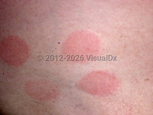
Allergic contact dermatitis - Oral Mucosal Lesion
See also in: Overview,Cellulitis DDx,External and Internal Eye,Anogenital,Hair and Scalp,Nail and Distal DigitAlerts and Notices
Important News & Links
Synopsis

Allergic contact dermatitis (ACD) is a type IV hypersensitivity reaction. In the oral mucosa, it can occur due to topical agents (including those applied to the lips), ingested agents, or implanted materials.
ACD reactions to dental implants most frequently present as lichenoid reactions that appear reticulate, atrophic, erosive, or plaque-like in appearance. This is often seen on the mucosa in contact with the culprit material. However, it can also present as a more diffuse stomatitis or burning / painful sensation for the patient (ie, burning mouth syndrome). One series of 22 patients with burning mouth syndrome who underwent patch testing found that 27% of patients had possible relevant sensitization to dental metals. Another study examined patients with various oral diseases (gingivitis, oral stomatitis, cheilitis, etc) and found that allergen patch test results were frequently positive not only for metal allergens but also for flavorings and preservatives.
Reactions to polyether impression materials used by dentists have also been noted.
Allergic contact cheilitis (flaking of the lips) can occur due to allergens in toothpastes, whereas perioral ACD has been reported by patients treated by dentists wearing protective rubber gloves.
Codes
L23.9 – Allergic contact dermatitis, unspecified cause
SNOMEDCT:
40275004 – Contact dermatitis
Look For
Subscription Required
Diagnostic Pearls
Subscription Required
Differential Diagnosis & Pitfalls

Subscription Required
Best Tests
Subscription Required
Management Pearls
Subscription Required
Therapy
Subscription Required
Drug Reaction Data
Subscription Required
References
Subscription Required
Last Updated:10/03/2024
 Patient Information for Allergic contact dermatitis - Oral Mucosal Lesion
- Improve treatment compliance
- Reduce after-hours questions
- Increase patient engagement and satisfaction
- Written in clear, easy-to-understand language. No confusing jargon.
- Available in English and Spanish
- Print out or email directly to your patient


