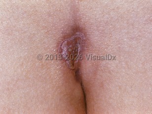
Pilonidal cyst
See also in: Cellulitis DDx,AnogenitalAlerts and Notices
Important News & Links
Synopsis

The spectrum of pilonidal disease includes asymptomatic sinuses and cysts as well as infected or inflamed cysts and localized abscess formation (also referred to as acute pilonidal disease). A pilonidal sinus results from a disruption of the epithelium in the gluteal fold overlying the coccyx, with formation of a small pit. Squamous epithelium gradually lines this cavity, which then plugs with hair or keratin, forming a cyst. Cysts can become inflamed or infected, and may rupture to form localized abscess formation. These abscesses contain a combination of skin and perineal flora. Chronic pilonidal disease is marked by draining sinus tracts or cysts with persistent drainage. Squamous cell carcinoma has been reported in chronic pilonidal cysts with incidence estimated to be around 0.1%; similar to squamous cell carcinomas that arise in chronic wounds, the biological behavior is thought to be more aggressive than squamous cell carcinomas that develop on sun-damaged skin.
Initially, pilonidal disease was believed to be congenital in nature and to represent a type of dermoid cyst. Current belief is that this is caused by excessive repetitive trauma to the sacrococcygeal region, illustrated by the prevalence of this problem among Jeep drivers in World War II (so called "Jeep disease").
Risk factors for pilonidal disease include male sex (adult), hirsutism, obesity, occupations requiring extended periods of sitting, and the presence of a deep natal cleft. Pilonidal cysts are more common in adolescence and young adulthood. Systemic signs and symptoms are rare.
"Barber's interdigital pilonidal sinus" refers to the reaction surrounding an entrapped cut or shaved hair, often seen in an interdigital space of a barber's hand. These cases represent foreign body granulomas rather than true cysts.
Codes
ICD10CM:
L05.01 – Pilonidal cyst with abscess
L05.91 – Pilonidal cyst without abscess
SNOMEDCT:
47639008 – Pilonidal cyst
L05.01 – Pilonidal cyst with abscess
L05.91 – Pilonidal cyst without abscess
SNOMEDCT:
47639008 – Pilonidal cyst
Look For
Subscription Required
Diagnostic Pearls
Subscription Required
Differential Diagnosis & Pitfalls

To perform a comparison, select diagnoses from the classic differential
Subscription Required
Best Tests
Subscription Required
Management Pearls
Subscription Required
Therapy
Subscription Required
References
Subscription Required
Last Reviewed:09/09/2021
Last Updated:09/09/2021
Last Updated:09/09/2021
 Patient Information for Pilonidal cyst
Premium Feature
VisualDx Patient Handouts
Available in the Elite package
- Improve treatment compliance
- Reduce after-hours questions
- Increase patient engagement and satisfaction
- Written in clear, easy-to-understand language. No confusing jargon.
- Available in English and Spanish
- Print out or email directly to your patient
Upgrade Today

Pilonidal cyst
See also in: Cellulitis DDx,Anogenital
