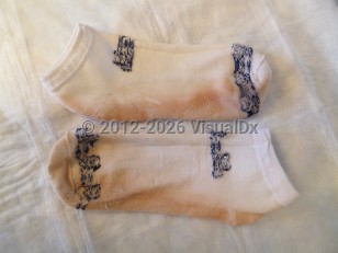
Chromhidrosis in Adult
Alerts and Notices
Important News & Links
Synopsis

Chromhidrosis may be further divided into apocrine and the much less common eccrine form. Not surprisingly, apocrine chromhidrosis is limited to apocrine gland-bearing areas such as the axilla, face, neck, and areola. Apocrine chromhidrosis is more commonly diagnosed in young adults. There is no predilection by sex. Common colors include yellow, green, blue, or black, and coloration is secondary to abnormal accumulation of lipofuscin within apocrine cells. Lipofuscin is a pigment that results from lysosomal digestion of lipids; why abnormal accumulation results in cases of chromhidrosis is not known. The oxidative state of lipofuscin determines the color observed. Typically, the pigment displays fluorescence at 360 nm, providing a useful diagnostic clue. Eccrine chromhidrosis is secondary to accumulation of water-soluble dyes, which a patient either ingests in foods containing pigment or dye-containing drugs, or is otherwise exposed to, within eccrine glands. For example, red eccrine chromhidrosis secondary to consumption of snack food colored with a red dye has been reported. Hyperbilirubinemia is another common cause of eccrine chromhidrosis, in which patients present with green-colored sweat. Eccrine chromhidrosis is more common on eccrine gland-bearing areas such as the hands and feet. In one small study, it was reported to be more commonly diagnosed in adult males, but the exact sex ratio is unclear given the rarity of this condition.
Pseudochromhidrosis, on the other hand, is often secondary to microbial overgrowth on the skin and accompanying pigment accumulation but may also be caused by other dyes contacting the skin, such as from clothing. In a frightening "epidemic" of cutaneous red spots on airline flight attendants, the source of the red dye turned out to be red ink on the demo life jackets. Bacterial species commonly implicated as causative agents include Corynebacterium, Pseudomonas, Bacillus, and Serratia species, all of which may produce pigment of various colors. Pseudochromhidrosis may occur on any cutaneous surface. It is typically diagnosed in young adults. There is no predilection by sex.
Codes
L75.1 – Chromhidrosis
SNOMEDCT:
26147006 – Chromhidrosis
Look For
Subscription Required
Diagnostic Pearls
Subscription Required
Differential Diagnosis & Pitfalls

Subscription Required
Best Tests
Subscription Required
Management Pearls
Subscription Required
Therapy
Subscription Required
Drug Reaction Data
Subscription Required
References
Subscription Required
Last Updated:05/08/2023

